
What is an echogenic intracardiac focus (fetal cardiac echogenic focus)?
An echogenic intracardiac focus is a hyperechogenic spot (aka bright spot) that is seen on a baby’s ultrasound in utero; the most common location of this “bright spot” is the left ventricle of the heart.
Like many topics about pregnancy, the echogenic intracardiac focus is surrounded by myths that scare the pregnant couple and that lack any scientific evidence or logic.
As you may know, the heart is composed of 4 small chambers; the upper two chambers are given the name: atria (plural of atrium), and the lower two are called ventricles. So, which chamber is the right or left one? The answer is in your first anatomy lecture below.
Anatomy 101
To be honest, it’s a bit tricky! You see, in medicine, we use a sense of direction that is based on the patient. What this means is that from the patient’s perspective, the heart is located on his left side and so it’s considered to be a left-sided organ by physicians.
Even though when the doctor is facing a patient, the heart of that patient is located on the right side from the doctor’s perspective.
In short, medicine and physicians see things from the patient’s perspective. which is a little cute if you think about it!
But enough about that for now. We’ll continue with a little lecture on radiology, but don’t let the word ‘lecture’ spook you out., it will all make sense by the end, I promise.
Radiology 101
In radiology, there are many machines and devices used to scan the human body to help with diagnoses and treatments.
Every machine has its particular properties and a different mechanism of action compared to another. For instance, an X-ray machine uses a beam of radioactive x-rays and shoots them at the patient to see what image is created.
During pregnancy; however, we have to be careful with what machine we use. You see, the fetus is undergoing a process called organogenesis (the formation of organs) which involves a lot of cellular divisions and DNA replication.
If you shoot X-ray beams at the mother’s uterus, the fetus will be vulnerable and the consequences are rather dire; ranging from simple harmless DNA mutations to complete malformations of the newborn.
For this reason, when a pregnant woman comes to the doctor’s office to check on her fetus, we don’t use X-rays machines or a CT scan which also involves using X-ray beams.
We are left with two options: MRIs and Ultrasound.
What do we use?
Ultrasound.
Why not use an MRI even though it’s clearer and more accurate?
One word: EXPENSIVE.
There you have it, you’re done with your first radiology lecture. If we keep this up, you’ll graduate from medical school in a week!
Now, let’s get back to the main topic of this article:
Echogenic intracardiac focus
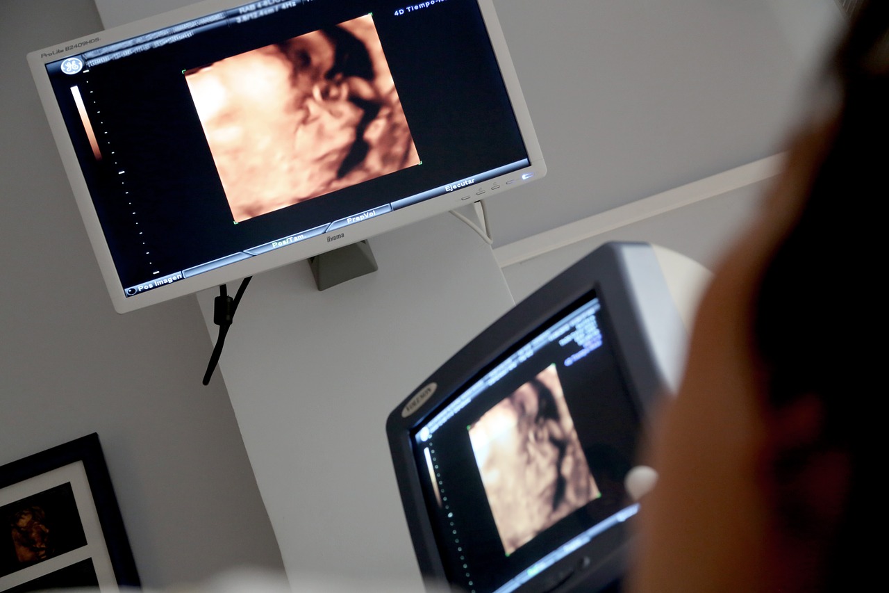
Like we said earlier, during pregnancy; the pregnant woman visits the doctor’s office multiple times for regular checkups.
Sometimes, during these checkups, the doctor will find a bright spot in the fetus’s heart. A spot that’s not supposed to be there.
The most common location is the left ventricle which now you know where it’s located. However, it can be found anywhere inside the heart, and on rare occasions, the bright spot isn’t an isolated finding. We can find multiple bright spots in the baby’s heart!
How common is an echogenic intracardiac focus?
Surprisingly, this finding is quite common. In fact, up to 20% or 1 in 5 babies are found to have an echogenic focus in the second or third trimester of the pregnancy.
Of course, this isn’t an exact number. You see, finding or missing an echogenic intracardiac focus depends on many factors, these include the baby’s position inside the uterus, the gestational age of the baby, the quality of the ultrasound machine, and the experience of the radiologist.
What causes an echogenic intracardiac focus?
You’ve probably have taken an X-ray before, right? The first thing you notice is that all your bones look white and bright on the X-ray; whereas, your muscles and soft tissues appear dark and invisible.
This is because your bones are mostly composed of minerals such as calcium and phosphate. These minerals are what we call radiopaque. Which is just a fancy word for a substance or a material that appears bright on a scan.
The same logic applies here; the heart is a muscle.
This muscle appears dim and dark on the ultrasound except for the valves and the septum. The first figure below shows how a normal heart looks on an ultrasound.
Sometimes; however, when a mineral such as calcium aggregates together. It forms this thick deposit of minerals that infiltrates inside the heart’s muscle. That spot is suddenly bright, and it’s what we call an echogenic (bright) intracardiac (inside the heart) focus (spot).
In other simple words, an echogenic intracardiac focus is nothing but the deposit of calcium and other minerals inside the heart’s myocardium (another fancy word for the heart’s muscle).
The Echogenic intracardiac focus and down syndrome
Usually, when an echogenic genic focus is seen on ultrasound; it’s not a big deal. We’ve already said that it’s seen in more than 20 percent of babies.
However, in certain situations and with high-risk mothers; it can be a problem. How?
Well, in 1995, a study was done by the Department of Obstetrics and Gynecology, Massachusetts General Hospital on 1334 pregnant women found that babies with a documented echogenic intracardiac focus had a significant risk of Down syndrome.
BUT! It’s rarely the case, most babies with an echogenic focus turn out to be perfectly fine with no diseases or malformation.
The risk only becomes really significant when there are other risk factors involved in the pregnant woman.
Risk factors for having a child with Down syndrome:
- Most importantly, advanced maternal age during the pregnancy.
- Having had one child with down syndrome before.
- Carrying a genetic dislocation for Down syndrome.
Is echogenic intracardiac focus dangerous?
The answer is no.
Although it can be associated with some genetic conditions such as Down syndrome, in most cases, it’s a fairly benign finding with no other implications.
Does echogenic intracardiac focus disappear?
As we’ve said earlier, most cases of echogenic intracardiac findings are seen in the second trimester.
The good news is that most of these cases disappear by the second half of the third trimester and have no consequences on the baby’s health and well-being.
Are there any other produces necessary?
If the pregnant woman has no other risk factors and the ultrasound shows no other signs of abnormal findings that suggest a genetic malformation, then no. No further exploration is needed and regular checkups should be scheduled normally.
However, if other risk factors are present. Your physician might order an amniocentesis, which is a procedure to take some amniotic fluid (the fluid that the baby floats in) and do karyotyping (chromosome analysis).
This procedure is done via the visual guidance of an ultrasound. The doctor will insert a thin needle into the patient’s abdomen until it reaches the amniotic sac where he/she can sample a few skin cells that physiologically shed from the baby. And then use this sample to analyze the number of chromosomes and the presence of genetic mutations.
Book recommendation
If you ask me what’s my favorite book to recommend to pregnant women? I’d immediately answer: What to Expect When You’re Expecting by Heidi Murkoff.
It’s a classic bestseller book with over 18 million copies sold. I’m sure many of you have heard of this book or read it already; it’s simply a MUST-READ for new and even experienced parents.
What I honestly love about it is that it is both informative and humorous, which is exactly what an expecting couple needs.
It basically covers every aspect of being pregnant from the psychological effect it’s going to have on the parents to the medical side of things (Zika virus, prenatal screening, and the safety of medications during pregnancy, as well as a brand-new section on postpartum birth control)
Current lifestyle trends are incorporated too: juice bars, raw diets, e-cigarettes, push presents, baby bump posting, the lowdown on omega-3 fatty acids, grass-fed and organic, health food fads, and GMOs).
Conclusion
Echogenic intracardiac focus is an ultrasound finding that is most commonly seen during the second and third trimester of pregnancy. It is associated with a higher risk of Down syndrome but in most cases, it’s just a benign finding.
Nevertheless, you should consult your primary care physician or Ob/Gyn doctor when you have any doubts or multiple risk factors of Trisomy 21 so you and your doctor can discuss the next best course of action.